PET, MRI show physical activity aids brain health
By Wayne Forrest, AuntMinnie.com staff writer
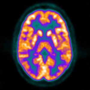
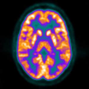
July 17, 2019 — PET (positron emission tomography) and MR (magnetic resonance) images show that older adults who are physically active have significantly less beta-amyloid accumulation and gray matter volume loss, which results in more stable cognitive skills, according to a study published online July 16 in JAMA Neurology.
The researchers also found that regular activity superseded the negative effects of vascular risk factors, such as hypertension, obesity, diabetes, and smoking, in preventing neurodegeneration and the potential onset of dementia.

“Together, these findings support interventions that target both physical activity and management of vascular risk factors as a means of delaying cognitive decline and neurodegeneration in preclinical Alzheimer’s disease,” wrote the researchers led by Jennifer Rabin, PhD, from Massachusetts General Hospital in Boston. “These lifestyle interventions could be coupled with anti-beta amyloid or anti-tau treatments when they become available.”
There have been many studies on how an active physical lifestyle might benefit one’s brain health by warding off the accumulation of beta amyloid and tau proteins, both of which are considered precursors to cognitive dysfunction and Alzheimer’s disease.
“However, it should be noted that most of these Alzheimer’s disease studies lacked assessment of beta-amyloid burden,” Rabin and colleagues noted. “In the present study, we examined whether baseline physical activity is protective against beta amyloid-related cognitive decline and neurodegeneration in a cohort of older adults who were clinically normal at baseline.”
The researchers recruited a total of 182 clinically normal older adults (mean age, 73.4 ± 6.2 years) from the Harvard Aging Brain Study, all of whom underwent PET imaging (ECAT Exact HR+ Siemens Healthineers) with carbon 11-labeled Pittsburgh Compound B (PiB) to obtain baseline beta-amyloid readings across the frontal, lateral temporal, parietal, and retrosplenial brain regions. All 182 subjects participated in the study through the fourth year of follow-up, while 96 people were still available by the seventh year of follow-up (mean follow-up, 6 years).
MRI scans to measure changes in gray matter volume were performed on a 3-tesla system (Magnetom Trio TIM, Siemens). At the three-year mark, 175 individuals had a follow-up MRI, and at the five-year mark, 98 subjects did.
At the start of the study, all participants had a global clinical dementia rating score of 0. Their mental acuity was monitored through the Preclinical Alzheimer Cognitive Composite, which includes a number of tests to determine memory and executive performance. Finally, to accurately measure their physical activity and eliminate any concerns of recall bias, the subjects wore pedometers for seven consecutive days while awake.
To quantify the results, the researchers calculated physical activity with changes in beta-amyloid levels, gray matter volume, and cognitive performance over the course of the follow-ups. They found greater physical activity was significantly associated with reduced beta amyloid-related cognitive decline (β coefficient, 0.03; p < 0.001) and reduced gray matter volume loss (β, 482.07; p = 0.002). In addition, vascular risk factors did not change the correlations between physical activity, slower beta amyloid-related cognitive decline (β, −0.04; p < 0.001), and reduced gray matter volume loss (β, −483.41; p = 0.01).
“Importantly, these associations remained significant after adjusting for vascular risk, supporting the view that the protective effect of physical activity on cognitive decline and neurodegeneration does not solely occur via mechanisms related to vascular risk,” Rabin and colleagues concluded.
Article from auntminnie.com
